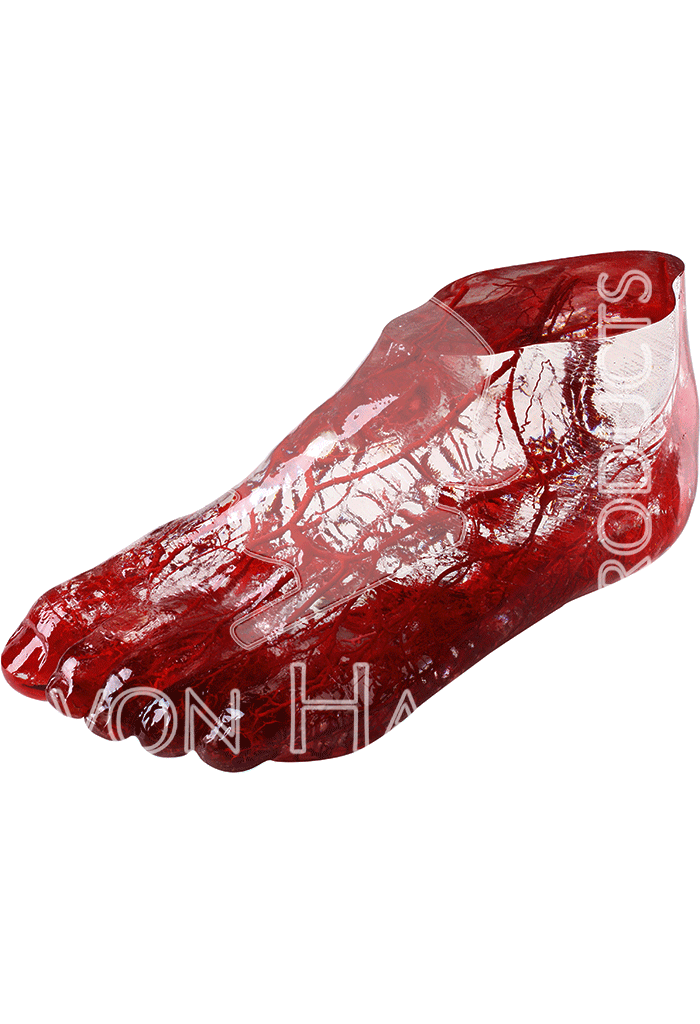
Description
Source Arteries
Blood supply of foot comes from three primary source arteries
Peroneal (fibular) artery
Posterior tibial artery
Anterior tibial artery
Peroneal (Fibular) Artery
Origin arises from the posterior tibial artery approximately 2.5 cm from its origin
Course in leg
pierces interosseous membrane ~ 5 cm above lateral malleolus as perforating branch and communicates with the anterior lateral malleolar artery
then passes down anterior to tibiofibular syndesmosis to anastomose with lateral tarsal artery
supplies the soleus, tibialis posterior, flexor hallicus longus, and peroneal muscles along its course
Branches at ankle
posterior lateral malleolar artery
communicating branch
Branches in foot
lateral calcaneal branch
terminal branch of the peroneal artery
provides perfusion to the lateral flap associated with a standard extensile approach to the calcaneus
Posterior Tibial Artery
Origin
largest of the two terminal branches of the popliteal artery
its most proximal part is referred to as the tibioperoneal trunk
Course in leg
it passes between the superficial and deep muscles of the posterior compartment of the lower leg
as it courses down the lower leg it becomes more medial and is palpable behind the medial malleolus
Branches at the ankle
posterior medial malleolar artery
communicating branch
artery of tarsal canal
dominant blood supply to the talar body
Branches in foot
beneath sustentaculum posterior tibial artery bifurcates into
lateral plantar arteries
branches
medial calcaneal branch (first branch)
is the major vascular supply to the heel pad
heel pad avulsions are severe injuries associated with high-energy trauma and often carry a poor prognosis because of the potential for heel pad necrosis
branches to adductor digiti minimi (second branch)
digital branch to fifth toe (third branch)
terminal branch
plantar branch (see below)
medial plantar arteries
branches
terminal branch
anastomoses with the first dorsal metatarsal branch of the dorsalis pedis artery
superficial digital branches join plantar metatarsal arteries of first three intermetatarsal spaces
Anterior Tibial Artery
Origin the other, smaller, terminal branch of the popliteal artery
Course in leg
descends anterior to the interosseous membrane and supplies the muscles of the anterior compartment of the lower leg
it becomes superficial at the ankle midway between the malleoli
supplies muscles of the anterior compartement of the lower leg
Branches at ankle
anterior medial malleolar artery
anterior lateral malleolar artery
Branches in foot
dorsalis pedis artery
a continuation of the anterior tibial artery in the foot
palpable over the dorsum of the foot just lateral to the extensor hallicus longus tendon
branches
arcuate (see below)
lateral tarsal
medial tarsal arteries
terminates at the first intermetatarsal space into
first dorsal metatarsal artery
deep plantar arch (see below)
Blood Supply to Distal Foot & Toes
Plantar archorigin
forms from the anastomosis of the lateral plantar artery and the dorsalis pedis artery
provides blood supply to plantar foot and toes
branches
plantar digital arteries
plantar metatarsal arteries
Arcuate artery
is a vascular arch that runs in the dorsal midfoot deep to the extensor tendons
gives off dorsal metatarsal arteries that run in the 2nd, 3rd and 4th intermetatarsal spaces.


Reviews
There are no reviews yet.